
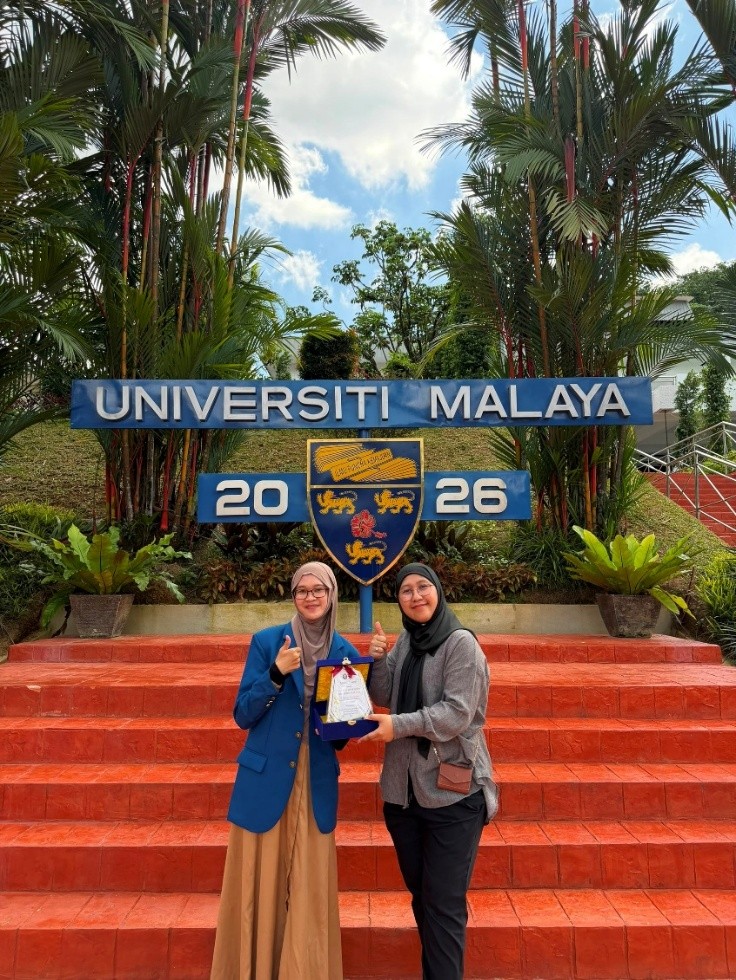
Kuala Lumpur, Malaysia — Gaining learning experience in an international academic environment plays a crucial role in developing student competence, particularly in broadening perspectives on advancements in science and technology. This was realized through the participation of Selvina Ariyanti, a Master’s student in Physics at the Faculty of Science and Mathematics (FSM), Universitas Diponegoro, in the Student Go International (SGI) – World Class University (WCU) Research Internship Delegation Program held at Universiti Malaya (UM), Kuala Lumpur, Malaysia, from August 2–8, 2026.
Throughout the program, the student engaged in various academic and research activities spanning nanotechnology, radiation physics, and medical physics. These activities not only provided an opportunity to explore research facilities but also highlighted the interconnectedness of material development, radiation technology, and their healthcare applications.
Expanding Nanotechnology Insights through Material Characterization
One of the hands-on research experiences took place at the Nanotechnology and Catalysis Research Centre (NANOCAT), Universiti Malaya. At this research center, the student was introduced to a wide range of facilities and instruments used for material research and characterization, particularly within the field of nanotechnology.
Various characterization techniques were covered, including X-Ray Diffraction (XRD), UV-Visible (UV-Vis) spectroscopy, Transmission Electron Microscopy (TEM), and zeta potential analysis. These techniques provided valuable insights into evaluating material properties across multiple dimensions, such as structure, optical traits, morphology, particle size, surface properties, and material stability.
The learning at NANOCAT directly aligns with her current research project—the synthesis of bismuth nanoparticles in chitosan solution as a candidate contrast agent for CT scans. A thorough understanding of material characterization is essential for evaluating synthesis outcomes and determining nanoparticle characteristics that could impact their effectiveness in medical imaging applications.
Exploring Radiation Technology and Research Instrumentation
The exploration of physics continued at the Radiation Laboratory, Department of Physics, Universiti Malaya. The laboratory environment offered the student firsthand exposure to advanced facilities and instrumentation related to radiation technology.
Instruments introduced during the visit included X-ray machines, GammaCell irradiators, High-Purity Germanium (HPGe) detectors, and Thermoluminescent Dosimeter (TLD) readers. In addition to touring the equipment, the student received detailed explanations regarding their working principles, functions, and research applications. This experience provided practical insights into utilizing radiation sources and measurement systems in a laboratory setting.
Observing the Application of Physics in Clinical Environments
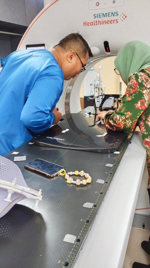
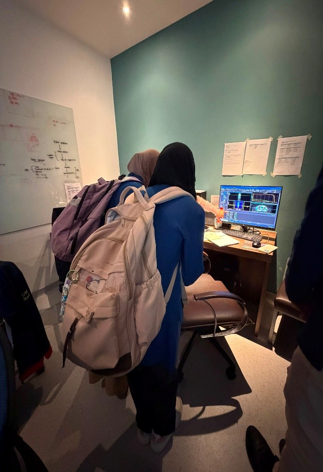
The next academic activity brought the student into a clinical environment through a visit to the Universiti Malaya Medical Centre (Pusat Perubatan Universiti Malaya – PPUM). This visit provided a firsthand look at how physics principles and medical technologies are practically applied in healthcare settings.
Among the technologies explored was Computed Tomography (CT-Scan), specifically focusing on the utilization of X-rays to generate diagnostic images. Through this activity, the student gained key insights into the role of imaging technology in clinical diagnosis and its direct connection to physics concepts studied in medical physics. Alongside CT scanning, the student was also introduced to radiotherapy, emphasizing the therapeutic application of radiation in patient care.
Learning within a clinical environment offered a perspective that complements laboratory experiences. While laboratory work introduces students to research protocols, instrumentation, and material characterization, the experience at PPUM demonstrated how advancements in physical technology are directly applied to meet real-world clinical needs.
Bridging Nanoparticle Research with CT-Scan Applications
The program concluded with academic discussions centered on research into synthesizing bismuth nanoparticles in chitosan solution as potential CT scan contrast agents. This discussion was highly relevant to the student’s ongoing research, addressing key experimental phases, critical material characteristics, and the potential of bismuth nanoparticles to enhance CT image contrast.
During these discussions, nanoparticle characterization emerged as a crucial step for evaluating synthesis outcomes. Comprehensive information on structure, morphology, particle size, optical properties, surface characteristics, and dispersion stability is necessary to fully understand material behavior. Relevant characterization techniques—such as XRD, spectroscopy, UV-Vis, TEM, and zeta potential analysis—were key components of the learning modules completed at NANOCAT.
The link between the properties of bismuth nanoparticles and their performance as contrast agents was also a major topic of discussion. In CT imaging, variations in X-ray attenuation between target materials and surrounding tissues govern image contrast. A contrast agent with higher X-ray attenuation capabilities can enhance this differential, allowing specific anatomical structures or target areas to be visualized with greater clarity.
Throughout the entire program, the student gained an integrated experience linking various domains of physics—from material characterization and radiation technology to medical imaging applications. This experience is expected to strengthen her academic and research competencies while expanding institutional connections with the academic community at Universiti Malaya to foster future research and academic collaborations.